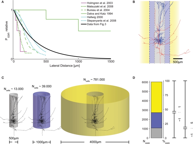
Figure 4.
Distribution of presynaptic cells within the cortical volume. (A) Graphical representation of connection probability as a function of lateral distance, comparing results from different studies. Methodological problems prevent direct numerical comparison. We, thus, normalized the maximum Pcon found in each study to unity and plotted one representative curve from each study into a single summary plot. Clearly, the length constant of the spatial decay of Pcon(d) derived from our data (black trace, cf. Figure 3H) fits well within the range reported by these previous studies. (B) Morphological reconstruction of a layer V pyramidal cell from a recording in an acute slice of 300 μm thickness with dendritic (blue) and axonal (red) arborizations. Following earlier work (Stepanyants et al., 2009), two definitions of locality can be derived from the neuronal morphology: either the volume covered by the dendrites (diameter of approximately 500 μm, gray), or, alternatively, by the dense axonal plexus around the somato-dendritic axis (diameter of approximately 1000 μm, blue). (C) The number of possible presynaptic partners (Ncells) increases substantially with distance, due to the quadratic increase of the volume covered by cylinders with increasing radius. This implies that the number of connected cells does not necessarily decrease with increasing distance, even if connection probability drops substantially. (D) To emphasize the consequences of the described distance dependence of Pcon for the total number of actually connected presynaptic cells within a certain distance, we calculated the numbers of these synaptically connected cells for the three different ranges depicted in (C). For all volumes, e used our exponential decay model with λ = 330 μm and P0 = 0.135 and, again, assumed a thickness of cortical gray matter of 1.3 mm. Surprisingly, even with a strong decay in Pcon(d) with increasing distance, the majority of presynaptic cells are located outside the local volume. Depending on the definition of locality, at least half of the synapses on each cell (local = diameter of 1000 μm), or more than 80% (local = diameter of 500 μm) originate from cells not considered to be within the local volume. The total number of presynaptic cells is slightly higher than what can be expected to be contained in a cylinder of 4000 μm diameter [cf. extent of the bar in (D) does not account for 100%].

