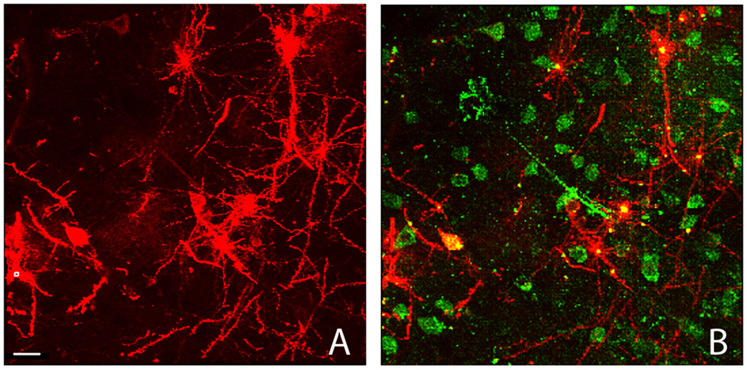
Figure 2.13.7.
Field view of DiI labeled tissue. A) Low power image of DiI labeled tissue slice. Scale bar = 20 µm. B) Over lay of DiI and immunofluorescent Hu staining, depicting the colocalization that is possible which enables the association of dendritic morphology with a neuronal phenotype (Copyright held by Robert Meisel).

