
Figure 2. Calcium mobilization is defective in Fyn null MCs but is unrelated to the depletion of intracellular calcium stores.
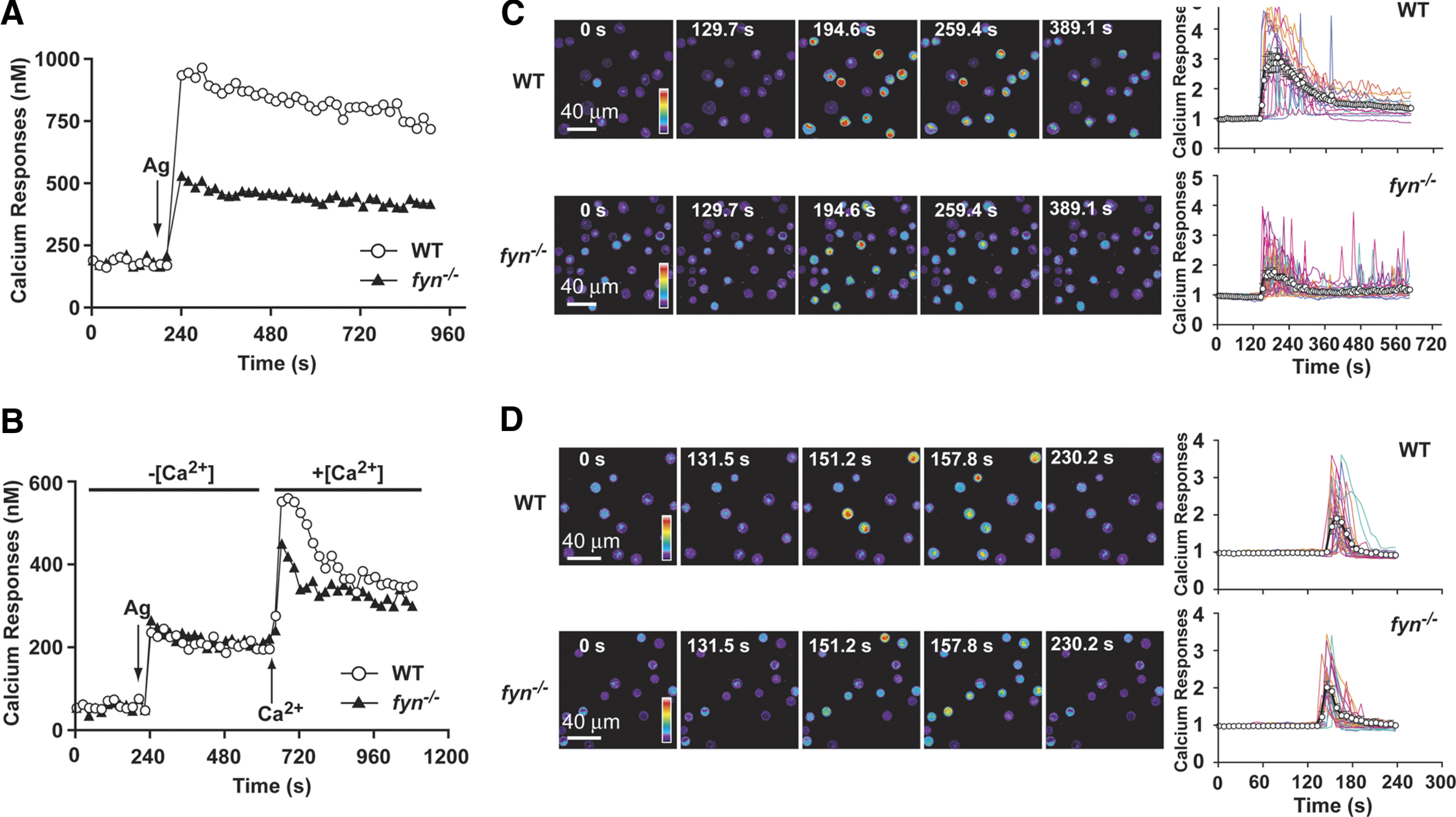
(A) IgE-sensitized WT or Fyn null MCs were loaded with Fura-2. MCs were stimulated with Ag (as indicated by arrow), and changes in the intracellular calcium concentration were monitored by fluorimetry for the indicated time. One representative of 4 experiments is shown. (B) WT and Fyn null MCs were treated in the same manner as above but were stimulated in the absence of extracellular Ca (–[Ca2+]), which was subsequently replenished to the extracellular medium (+[Ca2+]) as indicated. One representative of 3 experiments is shown. (C) Single cell analysis of calcium responses in WT and Fyn null MCs in calcium containing medium (+[Ca2+]). WT and Fyn null MCs were loaded with the calcium-binding Fluo-3 fluorescent dye and stimulated with Ag. Change in fluorescence intensity was monitored with time (as indicated). Reverse psuedo-colored images are used to distinguish the intensity of the individual cell response to the stimulus. (D) Single cell analysis of calcium responses in WT and Fyn null MCs in the absence of extracellular calcium. (C and D) Graphs were generated by capturing the fluorecense intensity of a single cell with time (128–326 cells were monitored in total). An averaged response is shown by the line with open circles.