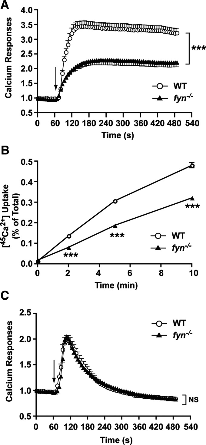
Figure 3. Thapsigargin stimulation of Fyn null MCs reveals normal depletion of intracellular calcium stores but impaired calcium influx.
(A) Thapsigargin (0.5 μM) induced a robust Ca2+ response from WT but not from Fyn null MCs in the presence of extracellular Ca2+. The arrow indicates the time of thapsigargin addition. Data are the mean ± se of all cells analyzed, and the statistical significance relative to WT (without treatment) was ***P < 0.001. One representative of 3 experiments is shown. (B) Thapsigargin stimulation of Fyn null MCs revealed a marked defect in 45Ca2+ uptake when compared with WT MCs. Data are the mean ± se of quadruplicate samples, and the statistical significance relative to WT (without treatment) was ***P < 0.001. One representative of 3 independent experiments is shown. (C) Thapsigargin-induced mobilization of intracellular Ca2+ in the absence of extracellular Ca2+ showed no difference in WT and Fyn null MCs. The arrow indicates the time of thapsigargin addition. Data are the mean ± se of all cells analyzed (42–51 cells were monitored in total). One representative of 3 experiments is shown. NS, Not significant.

