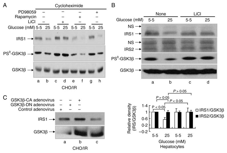
Figure 3.
Involvement of GSK3β in HG-induced IRS1 degradation. (A) CHO/IR cells were incubated in DMEM–0·25% BSA with cycloheximide (0·5 mM) and 5·5 or 25 mM glucose containing various inhibitors: LiCl (20 mM), rapamycin (200 nM), or PD98059 (0·1 mM) for 16 h. (B) Primary mouse hepatocytes were incubated in DMEM–10% FBS with 5·5 or 25 mM glucose for 22 h. In lanes c–d, LiCl (20 mM) was added to the medium and incubated for the last 8 h. (C) CHO/IR cells were infected with blank adenovirus or adenovirus containing the kinase-deficient GSK3β mutant or the constitutively active GSK3β mutant (200 MOI) for 20 h. After treatment(s), all cells were lysed in Laemmli sample buffer containing 0·1 M DTT. Lysates were processed for immunoblotting analysis as described in Materials and Methods. Results are representative of at least two separate experiments. Quantification of western blots (B) was done in the same way as described in Fig. 1. Data are expressed as means ± S.E.M., n = 3.

