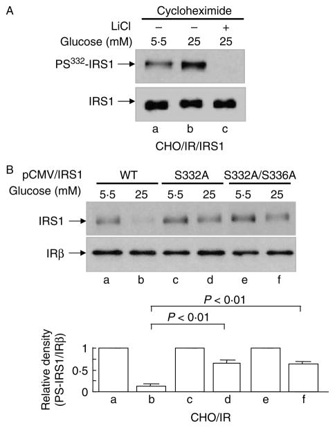
Figure 5.
Serine332 phosphorylation of IRS1 in HG-induced IRS1 degradation. (A) CHO/IR/IRS1 cells were incubated in DMEM–0·25% BSA containing cycloheximide (0·5 mM) and MG132 (25 μM) and 5·5 or 25 mM glucose in the presence or absence of LiCl (20 mM) for 24 h. (B) CHO/IR cells overexpressing wild-type IRS1, IRS1S332A, or IRS1S332/336A mutants were incubated in DMEM–0·25% BSA containing cycloheximide (0·5 mM) and 5·5 or 25 mM glucose for 16 h. After treatment(s), all cells were lysed in Laemmli sample buffer containing 0·1 M DTT. S332 phosphorylation of IRS1, IRS1 fprotein, and the β-subunit of insulin receptor in the lysates were detected by immunoblotting analysis using the corresponding antibodies as described in Materials and Methods. Results are representative of at least three separate experiments. Quantification of western blot (B) was done in the same way as described in Fig. 1. Data are expressed as means± S.E.M., n = 3.

