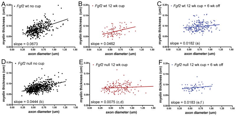
Figure 3.
Relationship of myelin thickness to axon diameter in Fgf2 null and wild type (wt) mice. Scatter plots of individual axons in the caudal corpus callosum for Fgf2 wt (A–C) and Fgf2 null (D–F) mice in non-treated (26 weeks) controls (A, D), demyelination after 12 weeks of cuprizone (cup) (B, E), and after 12 weeks of cup and 6 weeks on normal chow (C, F). (A–C) In Fgf2 wt mice, thinner myelin relative to axon diameter (indicative of remyelination) is demonstrated by the significantly reduced slope observed during the recovery period (a, Fgf2 wt, p < 0.05, no cup vs. 12 weeks cup + 6 weeks off). (D–F) In comparison with Fgf2 wt mice, Fgf2 null mice have thinner myelin prior to cup treatment (b, no cup p < 0.05, wt vs. null). Fgf2 null mice exhibit significantly reduced slopes from non-treated Fgf2 null mice after both 12 weeks of cup (c, p < 0.05, no cup vs. 12 weeks cup) and during the recovery period (e, p < 0.05, no cup vs. 12 weeks cup + 6 weeks off), indicating significant remyelination at the 12-week time point and continuing during the recovery period. Importantly, the slopes are also significantly different for the Fgf2 null mice vs. Fgf2 wt mice at 12 weeks of cup (d, wt vs. null, p < 0.05 at 12 weeks cup), indicating earlier remyelination in Fgf2 null mice than in wt mice. The slopes are not different for Fgf2 null vs. wt mice after the recovery period (f, wt vs. null, p > 0.05), indicating that the myelin thickness of remyelinated fibers is similar between genotypes even though Fgf2 null mice started with thinner myelin prior to cup treatment (b).

