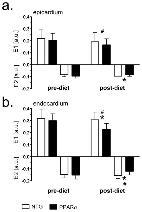
Figure 5.
Principal strains E1 and E2 before and after high-fat diet in midventricular septum of MHC-PPARα and NTG mice. a) epicardial values; b) endocardial values. Note reduced endocardial strains in MHC-PPARα hearts after high fat diet. * P<0.05, post-diet MHC-PPARα vs. post-diet NTG; # P<0.05, pre-diet MHC-PPARα vs. post-diet MHC-PPARα.

