
Figure 2.
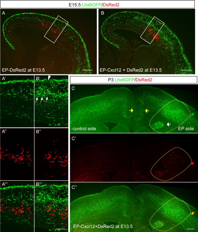
Ectopic Cxcl12 expression causes interneuron accumulation in vivo. A, B, Distribution of Lhx6-GFP+ cells at E15.5 was examined after in utero electroporation into the lateral cortex at E13.5 with either pCAG-DsRed2 alone (A) or together with pCAG-Cxcl12 (B). Ectopic Cxcl12 expression causes Lhx6-GFP+ cell accumulation, whereas DsRed2 expression has no apparent effect. A′ and B′ show the distribution of Lhx6-GFP+ cells at higher magnification. A″ and B″ show the distribution of DsRed2+ cells. A‴ and B‴ show the merged images. Scale bars: A, B, 200 μm; A′–A‴, B′–B‴, 100 μm. C–C″, Distribution of Lhx6-GFP+ cells at P3 was examined after in utero electroporation with pCAG-DsRed2 with pCAG-Cxcl12 at E13.5. There is notable accumulation of GFP+ cells around the ectopic site of expression of Cxcl12 (yellow oval) and a decrease in cell number in the medial cortex on the electroporated side (yellow arrows). Note as well that, where there was a small area of ectopic Cxcl12 expression in the SVZ (white arrow), there was a focal collection of GFP+ cells trapped in this area. EP, Electroporated. Scale bar, 300 μm.