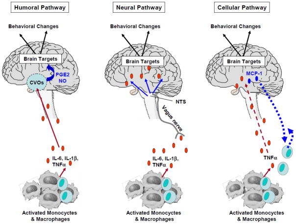
Figure 2. Communication Pathways from the Periphery to the Brain.
Different pathways by which cytokine signals access the brain have been identified.
- Humoral pathway: Pro-inflammatory cytokines released by activated monocytes and macrophages access the brain through leaky regions of the blood-brain barrier such as the choroid plexus and circumventricular organs (CVOs). Within the brain parenchyma, the activation of endothelial cells is responsible for the subsequent release of second messengers (e.g., prostaglandins [PGE2] and nitric oxide [NO]) that act on specific brain targets.
- Neural pathway: Pro-inflammatory cytokines released by activated monocytes and macrophages stimulate primary afferent nerve fibers in the vagus nerve. Sensory afferents of the vagus nerve relay information to brain areas through activation of the nucleus of the tractus solitarius (NTS) and area postrema.
- Cellular Pathway: A cellular pathway has been recently described by which pro-inflammatory cytokines, notably TNF-alpha, are able to stimulate microglia to produce monocyte chemoattractant protein-1 (MCP-1), which in turn is responsible for the recruitment of monocytes into the brain (D’ Mello et al., 2009).
Abbreviations: Interleukin-6: IL-6; interleukin-1β: IL-1β; tumor-necrosis factor: TNF

