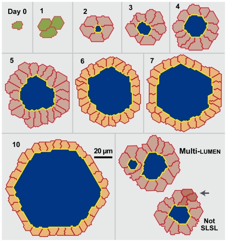
Figure 4. In silico MDCK analogue cyst cross sections.
Note that a regular hexagon in hexagonal space maps to a circle in continuous space. Images are from a single simulation run using parameter settings from Table 2. Cells are unpolarized (green), polarized (gray) or stabilized (orange). Cell-cell and cell-matrix borders are red; cell-lumen borders are yellow; lumens are blue. Lower right panel: shown is a multi-lumen cyst. Not SLSL: this single lumen cyst does not have a single layer of cells. The arrow indicates two cells not in contact with lumen.

