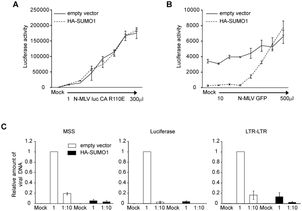
Figure 3. Characterization of SUMO-1 block of N-MLV infection in 293T cells.
A. The 293T empty vector control or HA-SUMO-1 cell lines were infected with increasing amounts of a mutant version of N-MLV luc in which amino acid 110 of the CA protein was mutated from arginine to glutamic acid (R110E). Luciferase activity was measured forty-eight hours post-infection. One representative experiment of three independent experiments is shown. B. The 293T empty vector control or HA-SUMO-1 cell lines were infected with increasing amounts of VSV-G-pseudotyped N-MLV expressing a GFP reporter, and four hours post-infection, cells were superinfected with a fixed amount of VSV-G-pseudotyped N-MLV luc. Luciferase activity was measured forty-eight hours post-infection. One representative experiment of four independent experiments is shown. C. The 293T empty vector control or HA-SUMO-1 overexpressing cell lines were infected with undiluted (1∶1) or 10-fold diluted (1∶10) VSV-G-pseudotyped N-MLV luc. Mock treated cells were included as a negative control. Total DNA was isolated twenty hours after infection, and the amount of viral DNA synthesized in the infected cells was measured by quantitative PCR. Primers specific for the minus-strand strong stop (MSS) DNA (left panel), Luciferase gene (middle panel) or LTR-LTR junction (right panel) were used. The values were normalized to GAPDH DNA and expressed as fold over empty vector. Error bars indicate standard deviation among triplicates in the same experiment (A and B) or 3 different experiments (C).

