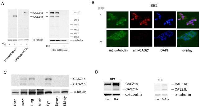
Figure 3. CASZ1a and CASZ1b are co-expressed and coordinately regulated at the protein level.
A. Anti-CASZ1 antibody is specific to CASZ1a and CASZ1b. Induction of CASZ1a and CASZ1b expression in the SY5YtetCASZ1a and SY5YtetCASZ1b clones by Tet at 24 hours was visualized by immunoblotting the cell lysates with anti-CASZ1 antibody (left); endogenous CASZ1a and CASZ1b expression in neuroblastoma cells was visualized by immunoblotting the BE2 cell lysates with anti-CASZ1 antibody, and the specificity of the antibody was demonstrated by pre-incubation of immunizing peptide and loss of CASZ protein recognition with immunoblotting analysis (right). B. Endogenous CASZ1a and CASZ1b are predominantly expressed in the nucleus of neuroblastoma cells. The nuclear localization of CASZ1a and CASZ1b was characterized by the co-localization of Alex 568-labeled goat anti-mouse IgG and DAPI-stained nucleus of BE2 cells (top), and the specificity of the antibody was demonstrated by the blocking effect of antigen peptide on antibody recognition with immunoblotting analysis (bottom). C. The co-expression of murine CASZ1a and CASZ1b protein in heart, lung and eye of P6 mouse was visualized by immunoblot analysis of tissue lysates using an anti-CASZ1 antibody. D. The simultaneous up-regulation of CASZ1a and CASZ1b protein by either RA or 5-Aza-dC in neuroblastoma cell lines was visualized by immunoblot analysis of the cell lysates using the anti-CASZ1 antibody.

