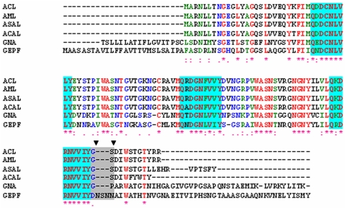
Figure 1. Alignment analysis of the deduced amino acid sequence of ASAL with closely related mannose binding homologues.
Sequence alignment among ASAL, Amorphophallus paeonifolius lectin (ACL), Arum maculatum lectin (AML), Allium cepa lectin (ACAL), Galanthus nivalis agglutinin (GNA) and Gastrodianin (GAFP). All identities and similarities are indicated by (*) and (:). Blue blocks and the grey box represent all mannose binding domains and the stretch of amino acids that differ in the monomer, respectively. The two residues at positions 98 and 99 (GNA numbering) are shown with two down triangles, where a trans-peptide bond is present in monomers instead of the cis-peptide bond found in all oligomers.

