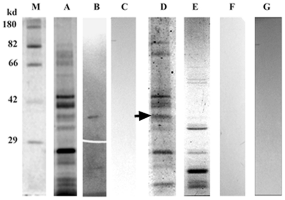
Figure 10. Identification and characterization of fungal receptor.
(A) Staining of the subproteome of R. solani using a Sypro ruby stain. (B) An approximately 37 kDa putative receptor was identified in a ligand blot assay of the membrane subproteome of R. solani with mASAL when probed with an anti-ASAL antibody. (C) Ligand blot analysis of the subproteome with mASAL pre-saturated with excess α- D mannose and probed with an anti-ASAL antibody showed an absence of signal (D) Glycospecific-staining of subproteome indicated the glycoprotein nature of the putative receptor (arrowhead denoting an approximately 37-kDa receptor) (E) Staining of deglycosidase-treated subproteome by Sypro ruby stain (F) Ligand blot analysis of de-glycosylated subproteome probed with an anti-ASAL antibody showed no signal (G) A ligand blot assay of a subproteome without mASAL as a negative control (M) represents a standard Protein molecular weight marker.

