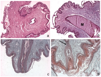
Figure 5. Nipples from nm23-M1−/− mammary glands are obstructed.
Sections from WT (a, c) and nm23-M1−/− (b, d) nipples were sectioned and stained by hematoxylin and eosin (a, b) or trichrom Masson (c, d). The general architecture of dermis and epidermis of the mutant nipple doesn't seem overly changed. However, close examination revealed that the final opening of the lactiferous canal is obstructed by epithelial cells in the mutant glands (arrow). M: milk. Original magnification X200 (a, b) X400 (c,d).

