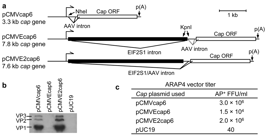
Figure 2. Intron-containing cap constructs.
(a) Schematic of intron-containing cap constructs. (b) Western blot of cell lysate of 293T cells transfected with 10 µg DNA using anti-AAV VP1+VP2+VP3 mouse monoclonal antibody (American Research Products). 10 µl of lysate was loaded per lane. (c) Crude AAV lysates were collected three days after transfection of 293T cells with 4 µg pARAP4, 4 µg pDGM6Δcap and 2 µg of a cap plasmid. Cells and medium were freeze/thawed three times, centrifuged at 1,000 × g for 10 min to remove cells and debris, and filtered (0.2 µm-pore-size). ARAP4 titers from one experiment were determined by infecting HTX cells and staining for AP+ foci after three days.

