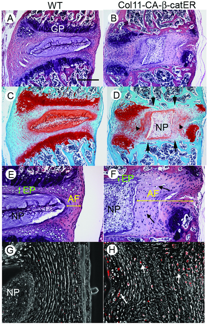
Figure 4.
Disorganization of the IVD structure in tamoxifen-treated Col11 CAβ-catER mouse. Col11 CAβ-catER mice (B, D, F, H and J) and their WT littermates (A, C, E, G and I) received seven daily injections of tamoxifen (200 µg/20 µl/day). Four weeks after the treatment, the vertebrae were dissected. The tissue sections were prepared and subjected to HE (A, B, E and F) or Safranin O (C and D) staining, immunostaining for anti-BrdU antibody (G and H). A bar represents 300 µm for A–D, 150 µm for E and F, and 75 µm for G and H.

