
Fig. 3.
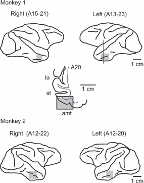
Recording positions. Activity of cells was recorded from the anterior part of the inferotemporal cortex, in the posterior/anterior range between 12 and 23 mm anterior to the ear bar position. The recordings were limited to the cortical extent on the ventrolateral surface of the brain, from the ventral lip of the superior temporal sulcus to the medial bank of the anterior middle temporal sulcus. The recording positions are shown by shading on the lateral views of the hemispheres. A ventrolateral part of the frontal section of the left hemisphere of Monkey 1 at anterior 20 is also shown in the middle. la, lateral fissure; st, superior temporal sulcus; amt, anterior middle temporal sulcus.