
Figure 6.
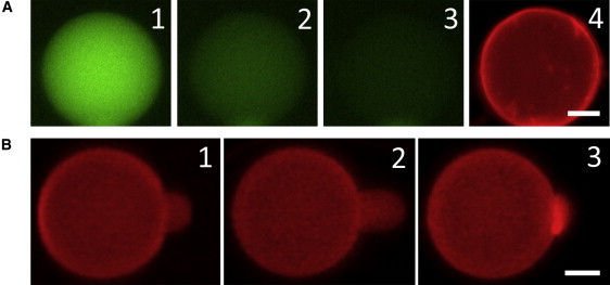
(A) GUV with red dye in the lipid and green dye in its content was exposed to 5 μM LL-37. Leakage occurred stochastically. 1) t = 0, right before leakage occurred. 2) t = 30 s. 3) t = 60 s. 4) t = 300 s (the GUV was still intact). Within 60 s, the leakage reduced the content dye intensity to ∼10% of the t = 0 value, whereas photobleaching decreased the intensity of a nonleaking GUV to ∼90%. Leakage was complete at t ∼ 200 s. (B) 1) An aspirated GUV was exposed to 0.5 μM LL-37. 2) The protrusion length initially increased, indicating a membrane area expansion without pore formation (the image shows the maximum protrusion). 3) After reaching the maximum, the protrusion length decreased, indicating pore formation in the membrane (see text). In nine runs of aspiration experiments, the average time to reach the maximum protrusion was ∼13 s and the average time to decrease to the original protrusion length (where ΔA/A = 0) was ∼10 s. Both scale bars = 10 μm.