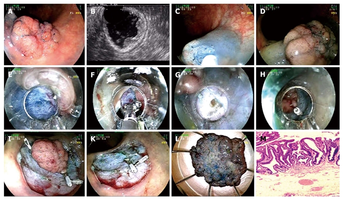
Figure 3.

Endoscopic submucosal dissection procedure for tubulovillous adenoma with high grade dysplasia at sigmoid colon. A: Adenoma at sigmoid colon; B: Endosonographic image of tubulovillous adenoma; C-D: Lifting the lesion; E: Cutting with endo-cut; F: Coagulation of submucosal vein with hemostatic forceps; G: Mini perforation during the procedure; H: Fixing perforation with hemoclip; I: Hemoclip application to control bleeding that occured after cutting the lesion circumferentially with endo-cut; K: Appearance of the mucosa after the lesion being extracted; L: Microscopic view of the lesion; M: Histology; tubulovillous adenoma with high grade dysplasia (HE × 40).
