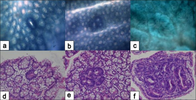
Figure 3.

Histopathology of early colonic neoplasms developed in AOM/DSS-treated mice. Single aberrant crypt (a, d); aberrant crypt focus - ACF (b, e); microadenoma (c, f). Methylene blue (a, b, c) and hematoxylin-eosin (d, e, f) stain. Original magnification, (a, b, c) ×10; (d, e, f) ×20. (Unpublished data).
