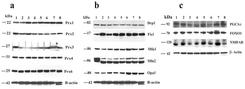
Figure 1.
Representative immunoblots of peroxiredoxins, mitochondrial proteins, and neuroprotective proteins in control N2a neurons, Aβ-incubated N2a cells, and N2a neurons treated with MitoQ and SS31 and then incubated with Aβ (n=3). Image A. Fifty μg of protein lysate was used from each cell sample; immunoblotting analysis was performed using antibodies of peroxiredoxins (1, 2, 3, 4, and 6). Image B. Drp1, Fis1, Mfn1, Mfn2, and Opa1. Image C. PGC1α, FOXO1, and NMDAR. Bottom panel shows immunoblotting of β-actin for equal loading. Lane 1 represents control N2a cells; lane 2, Aβ-incubated N2a cells; lane 3, resveratrol-treated N2a neurons; lane 4, N2a cells treated with resveratrol and then incubated with Aβ; lane 5, MitoQ-treated N2a cells; lane 6, N2a cells treated with MitoQ and then incubated with Aβ; lane 7, SS31-treated N2a cells; and lane 8, N2a cells treated with SS31 and then incubated with Aβ.

