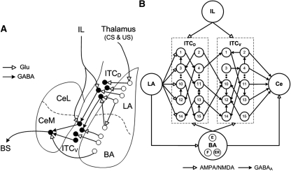
Figure 1.
(A) Scheme showing connectivity of the amygdala (adapted from Pare et al. 2004 and reprinted with permission from The American Physiological Society © 2004). LA receives thalamic inputs conveying information about the conditioned stimulus (CS) and unconditioned stimulus (US). LA projects to the basal nucleus (BA), and ITC neurons located dorsally (ITCD), which in turn project to ITC cells located more ventrally (ITCV). ITCV cells contribute GABAergic projections to CeM (central medial nucleus). The BA sends excitatory inputs to both ITCD and ITCV cells, and to CeM. IL also projects to both ITCD and ITCV cells. CeM projects to brainstem structures mediating fear responses. (BS) Brainstem; (Glu) glutamate. (B) Structure of the model ITC network with 15 neurons each in ITCD and ITCV clusters. Each ITC neuron inhibits three randomly selected neurons in the same cluster (only one projection per neuron is shown in the figure). Each ITCD neuron also inhibits three randomly selected ITCV neurons (e.g., ITCD2 inhibits ITCV10). For clarity and illustration purpose, the figure only shows partial connectivity, which may not be the actual connectivity used in the model (see Supplementary Table S3). The network has five Ce output neurons that receive excitatory inputs from BA, and inhibitory inputs from ITCV neurons. ITCD and ITCV: Neurons 1–5 are of type A neurons (with facilitating output synapses); neurons 6–10 are of type B (with depressing output synapses); and neurons 11–15 are of type C neurons (with constant synapses). See the Materials and Methods section entitled “Presynaptic release probability” for description of facilitating and depressing ITC synapses.

