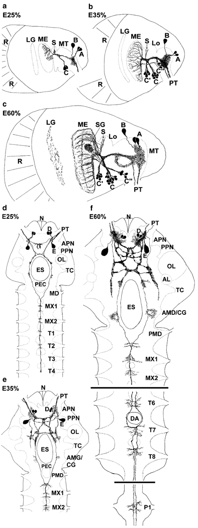
Fig. 1.
Representations summarising the PDHir structures in embryonic lobster eyestalks (a–c) and median brains and ventral nerve cords (d–f) from 25% to 60% of embryonic development (E25%–E60%). Groups of neuronal somata are denoted by letters A–E (AL accessory lobe, AMD/CG anterior part of the mandibular neuromere, which is termed commissural ganglion in adult crustaceans, APN anterior protocerebral neuropil, DA descending artery, ES oesophageal foramen, LG lamina ganglionaris, Lo lobula, ME medulla externa, MT medulla terminalis, MX1 neuromeres of maxilla 1, MX2 neuromeres of maxilla 2, N nauplius eye, OL olfactory lobe, P1 pleon neuromere 1, PEC postoesophageal commissure, PMD posterior part of the mandibular neuromere, PPN posterior protocerebral neuropil, PT protocerebral tract, R retina, S medulla satellite neuropil, SG sinus gland, T1–8 thoracic neuromeres 1–8, TC tritocerebrum)

