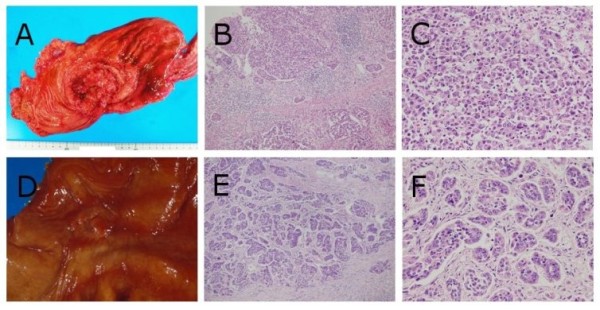
Figure 1.

Macroscopic and histopathological findings of gastric carcinomas in case 1 and 2. Case 1 (A) The ulcerated tumor with relatively demarcated and raised margins (Borrmann's type 3 tumor) was located in the gastric body. (B and C) Poorly differentiated adenocarcinoma cells diffusely proliferated (hematoxylin-eosin (HE) double stain, × 100, × 400, respectively). Case 2 (D) The ulcerated tumor with demarcated and slightly raised margins (Borrmann's type 2 tumor) was located in the gastric body. (E and F) Poorly differentiated adenocarcinoma cells were arranged in small nests or trabecular pattern surrounded by fibrous stroma (HE double stain, × 100, × 400, respectively).
