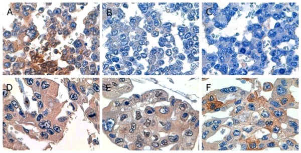
Figure 6.

Photomicrographs showing representative immunohistochemistry. (A, B, and C) Gastric carcinoma cells in case 1 showed positive reactivity for tissue factor and negative reactivity for vascular endothelial growth factor and osteopontin (× 1,000). (D, E, and F) Gastric carcinoma cells in case 6 showed positive reactivity for tissue factor, vascular endothelial growth factor, and osteopontin (× 1,000).
