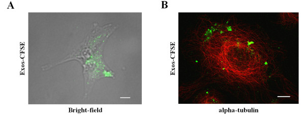
Figure 1.
Uptake of SKOV3 exosomes by SKOV3 cells. (A) SKOV3 cells were incubated with Exos-CFSE (20 μg protein; green) for 4 h and were visualized in bright-field merged with fluorescence microscopy. Scale bar = 20 μm. (B) Detection of Exos-CFSE (green) and alpha-tubulin (red) by confocal immunofluorescence microscopy. Scale bar = 10 μm.

