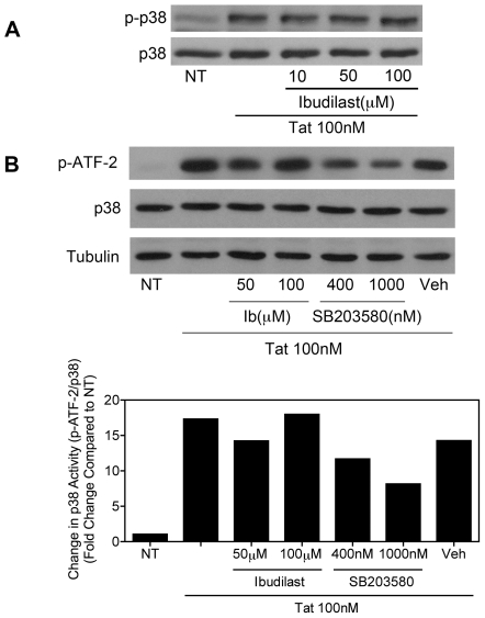
Figure 3. Ibudilast does not inhibit Tat-induced activation of p38 MAP kinase.
A, BV-2 cells (1.2×10⁵) were left untreated (NT) or were treated with Tat (100 nM) for 15 min. with or without pre-treatment for 5 min. with increasing concentrations of ibudilast, as indicated. Whole cell lysates were subjected to immunoblot analysis using either phospho-p38 MAPK-specific (upper panel) or p38 MAPK-specific (lower panel) antibodies. The results of a single representative experiment are shown; three total experiments were performed. B, BV-2 cells (2.4×10⁵) were left untreated (NT) or were treated with Tat (100 nM) for 20 min. with or without pre-treatment for 10 min. with increasing concentrations of ibudilast or the p38 MAP kinase inhibitor, SB203580, as indicated. Whole cell lysates were subjected to immunoprecipitation with an immobilized phospho-p38 MAP kinase antibody. Precipitated p38 was incubated with an ATF-2 fusion protein substrate. Phosphorylated ATF-2 levels were determined by immunoblot analysis with a phospho-ATF-2-specific antibody (top panel). Whole cell lysate supernatants collected after immunoprecipitation were subjected to immunoblot analysis using p38-specific (middle panel) or α-Tubulin-specific (lower panel) antibodies. Protein levels were quantified using ImageJ software (bottom graph). Phospho-ATF-2 levels were normalized to total p38 MAPK levels and fold change compared to NT was calculated. The results of a single representative experiment are shown; two total experiments were performed.

