
Figure 6. Electrophoretic Mobility Shift Assays with CtrA and SciP.
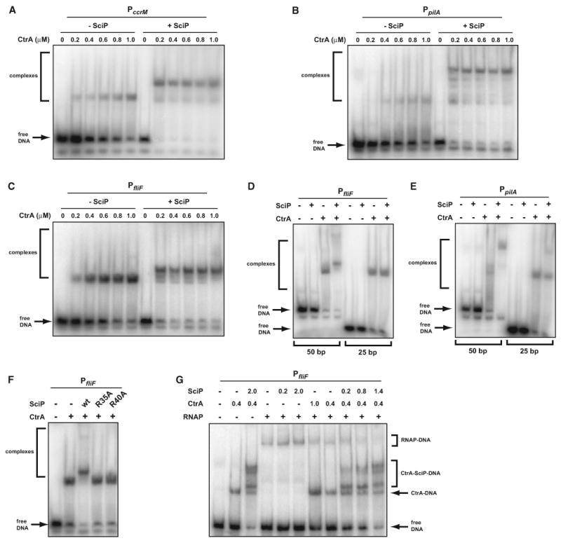
(A–C) CtrAP∼ binding to a 50 bp fragment of the (A) ccrM, (B) pilA, or (C) fliF promoters, each containing a CtrA-binding site. SciP, if present, was added at a final concentration of 1 μM. The concentration of CtrA in each lane is indicated.
(D and E) CtrA∼P binding to 25 bp fragments of the fliF and pilA promoters compared to the 50 bp probes also used in (B) and (C). CtrA was present at 1 μM and SciP at 1 μM.
(F) Binding of CtrA∼P to the fliF promoter in the presence of wild-type SciP or the mutant indicated. When included, CtrA was present at 1 μM and SciP at 1 μM.
(G) CtrA∼P and SciP prevent RNAP from binding the fliF promoter. CtrA∼P and SciP were added at the concentrations indicated. TAP-tagged RNAP was included, where indicated, at a final concentration of 50 nM (see the Experimental Procedures for details on RNAP preparation). (A)–(F) and (G) use 6% or 5%, respectively, polyacrylamide gels for separation.