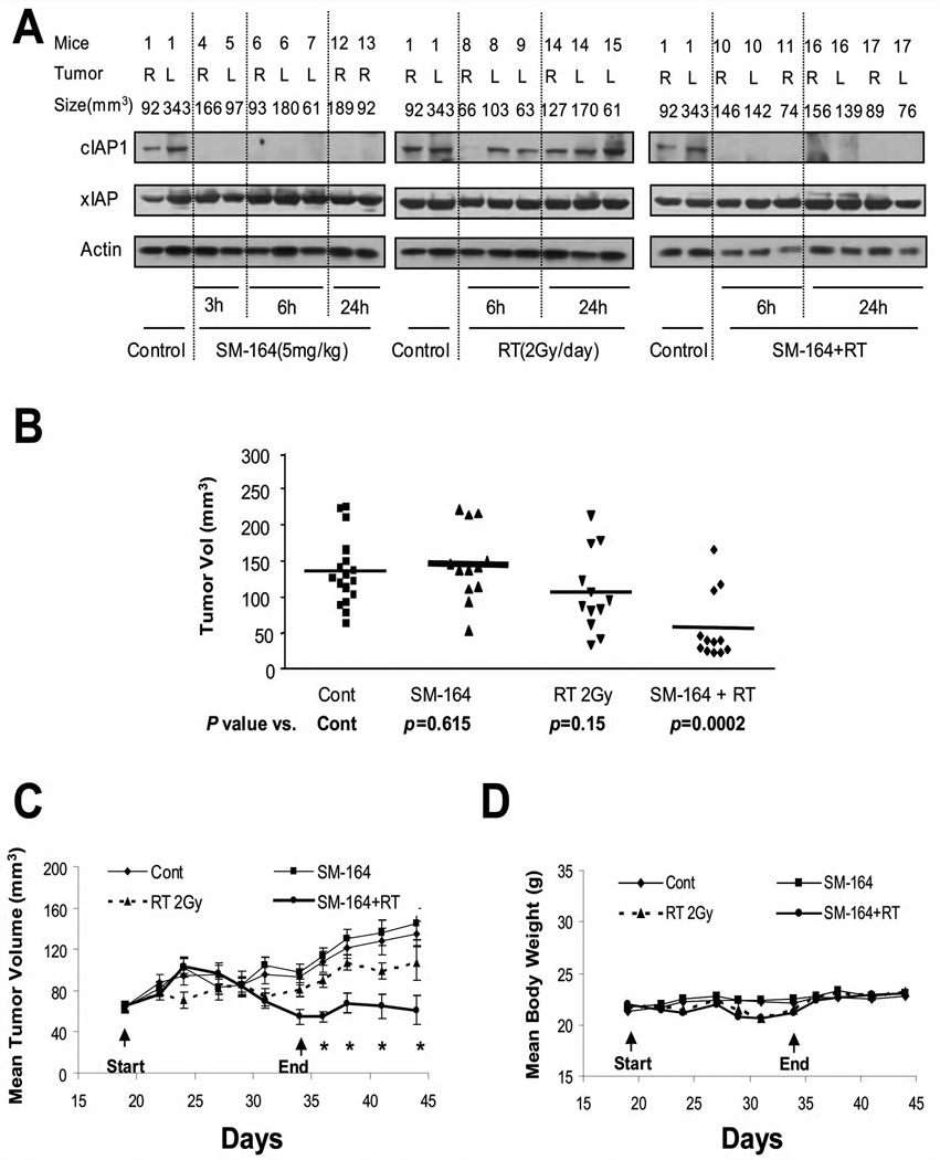
Figure 6. SM-164 radiosensitization in UMSCC-1 in vivo xenograft tumor model.
(A) SM-164 eliminated cIAP-1 in tumor tissues: In experiment one, five million UMSCC-1 cells were inoculated s.c in both flank sides (R, right; L, left) of nude mice. The mice were randomized when tumor size reached 70 mm3 at 18 days after inoculation and were left untreated or for a single treatment with SM-164, radiation, or drug-radiation combination. Tumor tissues were harvested at indicated time points for immunoblotting. (B &C) In vivo radiosensitizing activity of SM-164 in the UMSCC-1 xenograft model: In experiment 2, five million UMSCC-1 cells were inoculated s.c in both flank sides of nude mice. The treatment started 18 days after inoculation when tumor size reached 70 mm3, followed by tumor growth measurement. Student’s t test was used to compare each treatment group with the control group. Shown are the results on day 44 (mean ± SEM, * indicates p<0.05) (B), and tumor growth curve (C). (D). SM-164 is well tolerated by mice: Body weight was measured during the treatment and thereafter and plotted (mean ± SEM).

