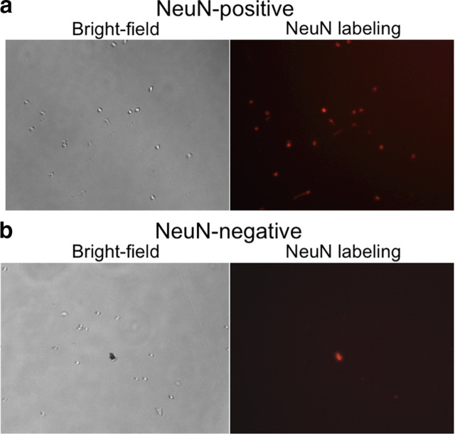
Figure 2.
Microscope photos of FACS-purified cells and debris. a, b, Bright-field and fluorescence images of NeuN-positive (neural) (a) and NeuN-negative (glial) (b) cells after fluorescence-activated cell sorting. All cells are round and devoid of processes. NeuN labeling is red. In b, there is a piece of debris that is autofluorescent (shows up in every fluorescent light channel).

