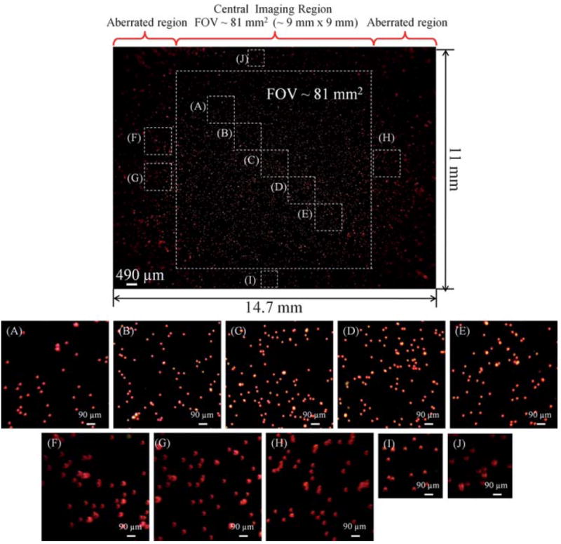
Fig. 2.

Imaging performance of the cell-phone fluorescent microscope shown in Fig. 1 is demonstrated using fluorescent beads (10 μm diameter; excitation/emission: 580 nm/605 nm). The central field-of-view of each cell-phone image is ~8l mm2, which exhibits a decent imaging performance. The edges of the image, which lie outside of this central region exhibit aberrations, and therefore are not included in the reported field-of-view. For counting purposes, however, those aberrated regions could still be useful despite their poorer image quality. Note that all the scale bars in zoomed frames (A–J) have the same length.
