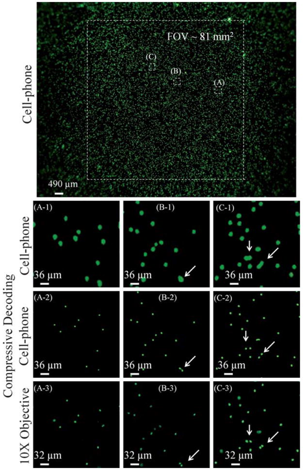
Fig. 4.

Imaging performance of our cell-phone fluorescent microscope is demonstrated using labeled white blood cells. Microscope objective (10×, NA = 0.25) images of the same samples, acquired with a conventional fluorescent microscope, are also provided for comparison purposes. White arrows point to cells that can be resolved using compressive decoding, which further demonstrate our improved resolving power similar to Fig. 3. Note that because the samples were suspended in a solution, their relative orientations might be slightly shifted in microscope comparison images, as a result of which the FOV and the scale-bars of the microscope images are slightly different when compared to the cell-phone images.
