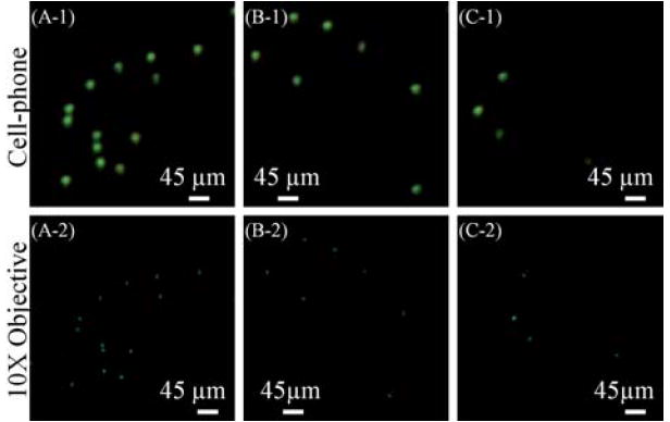
Fig. 5.

(Top) Giardia Lamblia cysts that are imaged using the fluorescent cell-phone microscope of Fig. 1. (Bottom) Microscope objective (10×, NA = 0.25) images of the same samples are also provided for comparison purposes. Note that because the samples were suspended in a solution, their relative orientations might be slightly shifted in the microscope comparison images. In (B-2) and (C-2) there are 2 dead-pixels at the microscope images which do not show up in our cell-phone images.
