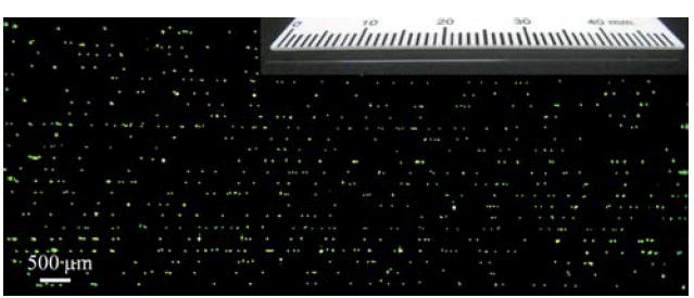
Fig. 6.

Fluorescent samples can also be imaged within micro-capillaries using our cell-phone based fluorescent microscope. In this case, simple capillary action is sufficient to load the specimen into a capillary tube. Each capillary, when loaded with the sample solution, acts as a wave-guide for pump photons, such that efficient excitation of the samples could be achieved as illustrated in this figure for 10 μm fluorescent beads that were loaded into several capillary tubes in parallel. The inset figure at the top corner illustrates one of the capillaries used in this work (100 μm inner diameter; 170 μm outer diameter). For further information please refer to Supplementary Fig. 1.†
