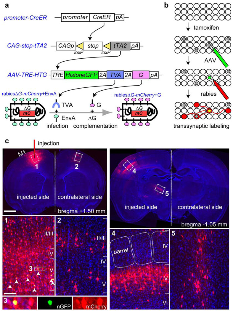
Figure 1. Genetic control of rabies-mediated neural circuit tracing.
a-b, Schematic representation of the methodology used to control the location, number and type of starter cells for RV-mediated transsynaptic labeling. tTA2 is expressed in a small subset of CreER(+) cells (grey nuclei in b). tTA2 activates an AAV-delivered transgene to express: 1) a histone-GFP marker to label the nuclei of starter cells in green, 2) EnvA receptor (TVA) to enable subsequent infection by EnvA-pseudotyped RV (rabies ΔG-mCherry+EnvA), and 3) rabies glycoprotein (G) to initiate transsynaptic labeling. c, Top left, a 60-µm coronal section that includes the injection site in the motor cortex (M1). Cells labeled with both histone-GFP (n-GFP) and mCherry (arrowheads in c1, magnified in c3) can be distinguished from cells labeled with mCherry alone, which are found near the injection site (c1), in the contralateral motor cortex (c2), in the somatosensory barrel cortex (top right; magnified in c4), and in the motor-specific ventrolateral nucleus of the thalamus (c5). Scale bars, 1 mm for low-magnification images on top, 100 µm for high-magnification images at the bottom.

