Figure 2.


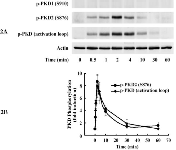
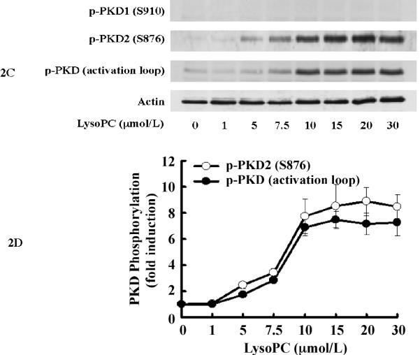
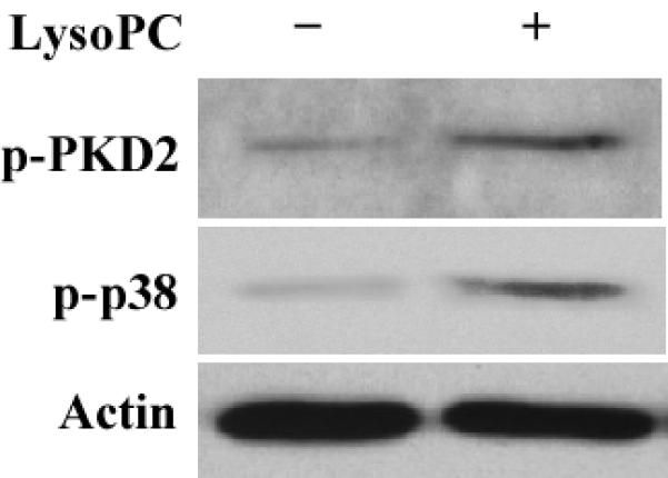
LysoPC induces PKD2 activation in THP-1 cells and monocytes. A, Time course (Western analysis) with LysoPC (15 μmol/L) used to stimulate THP1 cells. Cells were cultured in growth medium for 2 days and then incubated with or without lysoPC (15 μmol/L) for the indicated times. Phospho-specific antibodies against p-PKD1 (S910), p-PKD2 (S876), and p-PKD (activation loop) were used. Actin was used to assess protein loading. B, Time-dependent PKD phosphorylation was quantified by densitometry. C, LysoPC concentration dependence of PKD activation at 2 min in THP1 cells (Western analysis). D, Concentration-dependent PKD2 activation was quantified by densitometry. Data are means ± SE from 4 independent experiments. E, lysoPC (15 μmol/L) induces phosphorylation of PKD2 and p38 MAPK in monocytes (2 min).
