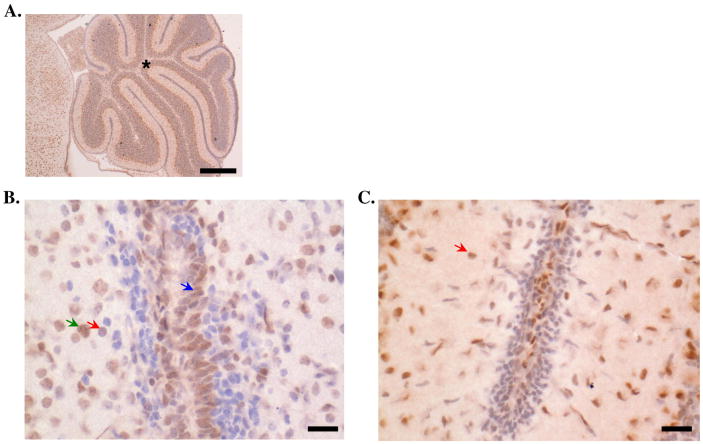
Figure 3. Glucocorticoid receptor expression in brain tissue.
Expression of the stress hormone receptor, GR, was evaluated in mouse brain tissue via immunohistochemistry. Asterisk is located in the granular layer of the cerebellum. GR expression (indicated by positive cytoplasmic staining) was seen in several cell types identified based on morphology, including microglia (red arrows; approximately 8-10 μm in diameter, round, hyperchromatic cells with no appreciable cytoplasm), astrocytes (green arrows; approximately 10-20 μm in diameter, large, vesiculate nuclei with distinct nucleoli and indistinct cytoplasm) and granule cells of the external granular layer (blue arrows; located within the inner and outer granular layers of the cerebellum, approximately 8-10 μm in diameter, round, hyperchromatic cells with scant cytoplasm). Micrographs of cerebellum at 20× (scale bar=300 μm, (A) and 400× (scale bar=50 μm, (B, C) magnification are shown. In Panel C, several microglia cells were identified with cytoplasmic GR staining. Micrographs shown are representative of 5 experiments.

