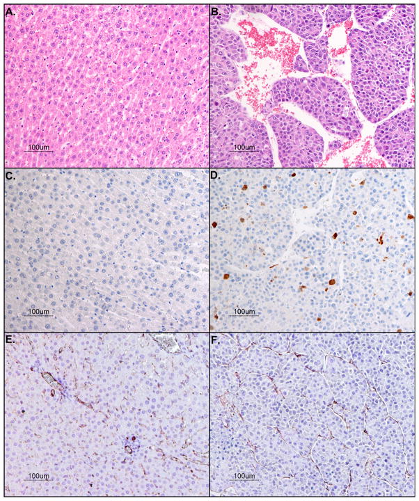
Figure 4.
Histopathologic analysis with hematoxylin and eosin stains of rat liver bearing hepatocellular carcinoma [A] normal liver and [B] tumor (20-fold magnification. Photomicrograph of anti-Caspase-3 positive stain for apoptotic cells [C] normal liver and [D] tumor (20-fold magnification). Immunostain of endothelial cells lining vessel walls with anti-CD31 (brown) and counterstained with hematoxylin for cell nuclei (blue) [E] normal liver and [F] tumor (20-fold magnification).

