
Figure 1.
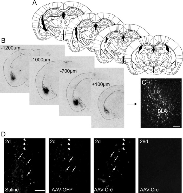
rAAV-Cre-induced expression of Cre mRNA is confined to the BLA. A, Schematic drawing from the mouse brain atlas by Paxinos and Franklin (2001) corresponding to sections containing the BLA. B, C, Autoradiographs (B) and a high-magnification dark-field photomicrograph of a photoemulsion-dipped section (C) showing strong but site-restricted expression of Cre mRNA after rAAV-Cre injection into the BLA. D, Fluoro-Jade C labeling of the injection site 2 d after injection with 1 μl of saline, rAAV-GFP, or rAAV-Cre and 28 d after rAAV-Cre injection. Note that the labeling was restricted to a small number of neurons (arrows) and similar for all three conditions after 2 d. No signs of degeneration were seen 28 d after vector injection. Arrowheads indicate the location of the needle tract. Scale bars: B, 1 mm; C, D, 500 μm.