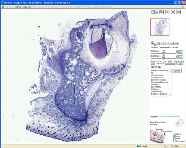
Figure 1.
Screenshot of WebMicroscope plug-in viewer. This is screen shot of the WebMicroscope for the oral pathology course. Using the navigational tools at the top of the right frame, student can manipulate the virtual slide through five levels of magnification, starting at the level of the whole mount, as well as click and drag the slide in an x-y axis through the entire surface of the slide, at any magnification. In the lower frame is the text of the laboratory syllabus. A view of early bell stage of odontogenesis is illustrated.

