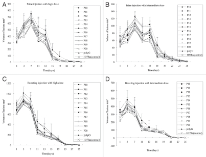
Figure 3.
Pathologic changes of rabbit skin lesions produced by different H37Ra complemented strains after prime and boosting immunity. Each point represents the mean with its standard error from four injection sites. Pathologic changes of the primary injection with high dose (A) and intermediate dose (B). High dose (5 × 10⁷ CFU) of bacteria induced granuloma 1–2 days after injection, while 1–5 days for intermediate dose (5 × 10⁶ CFU), and the injected area formed inflammation with edema. Inflammation reached peak about 5–7 days for high dose and 10–13 days for intermediate dose, then liquefied. At 35 days, the lesions epithelialized and healed. Pathologic changes of the boosting infection with high dose (C) and intermediate dose (D). The inflammation of high dose reached peak about 2–3 days and the intermediate dose need 3–7 days after last injection, shortly after this, the lesions liquefied and discharged its contents. These lesions healed at about 35 days. The tubercles caused by P10 was the largest, followed by P19, P17, P15, P18, P14 and others (*p < 0.05 in Table 3), while P14 and P18 induced the most severe liquefaction.

