
Figure 1.
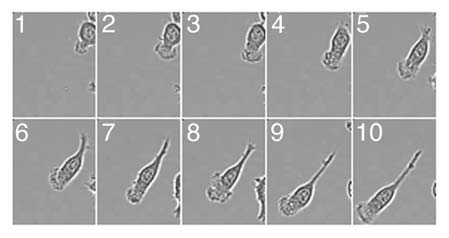
Dynamics of the lamellipodia formation in migrating macrophages. Macrophages plated on fibronectin coated glass bottom dishes (MatTek) were stimulated with 100 nM fMLP and imaged at 37°C in CO2 independent media. Movie frame rates were captured every 5 min over 3 h. Images were captured using Metamorph V67.1 software (Molecular Devices, Sunnyvale, CA) and edited using ImageJ. Selected frames are shown.