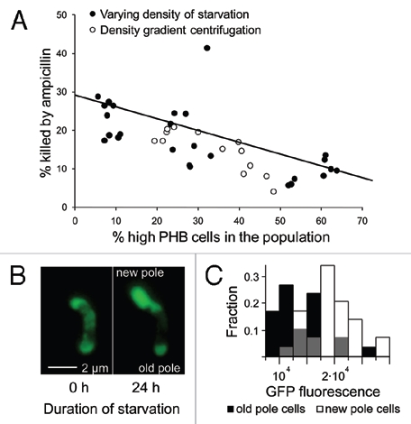
Figure 1.

High-PHB old-pole cells enter a persistent state. (A) Populations of Sinorhizobium meliloti that varied in the percentage of high vs. low-PH B cells were generated via density gradient centrifugation or by starving rhizobia at varying densities. These rhizobia were then challenged with ampicillin and assayed for mortality (PI or YO-PRO-1 uptake) by flow cytometry. Populations containing a greater fraction of high-PHB cells were less susceptible to ampicillin. There was no significant difference in linear regression slopes (t = 0.8, p = 0.43, n = 41, ANCOVA) or intercepts (t = 1, p = 0.32, n = 41, ANCOVA) for the two methods of manipulating the frequency of high-PH B cells, so a single linear regression was fit to the data. High-PH B old pole cells are less GFP fluorescent (B and C), indicating lower metabolic activity. (B) An initially high-PH B cell dividing during starvation produces a bright, GFP fluorescent new-pole cell and a dim old-pole cell. To increase brightness and lessen background fluorescence, this image was transformed with the ‘Colors; Levels’ command in GIMP v. 2.6. Both left and right panels were transformed identically. This transformation made the cells easier to see and did not qualitatively affect the image. (C) After 24 hours of starvation, GFP fluorescence intensity is 50.8% higher in new-pole cells (t = 5.29, n = 60, p < 0.0001, two-sided t-test). Gray bars represent overlap in new and old-pole distributions. Brightness units are average color depth (ranging from 0–65536) of the green channel from a 16-bit color fluorescence image, higher values indicate greater brightness.
