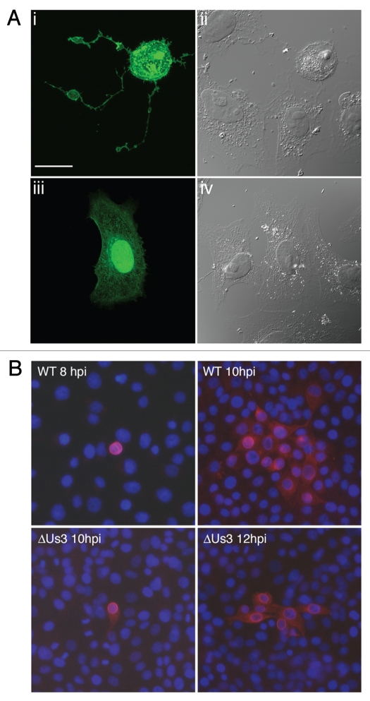
Figure 1.
PRV Us3 induces filamentous process (FP) formation and influences cell-to-cell spread of PRV. (A) FPs induced by PRV Us3. Vero cells were transfected with plasmid encoding a PRV Us3-GFP fusion protein (i and ii) or GFP (iii and iv) and fixed at 24 hours post transfection. Fixed specimens were examined by confocal microscopy. Note the long FPs extending from the Us3-transfected cell and also that the Us3-transfected cell has a rounded morphology indicative of actin stress fibre breakdown. Scale bar is 20 µm. (B) Spread of PRV in the absence of Us3. PK15 cell monolayers were infected with wild type (WT) or Us3 null (ΔUs3) PRV at a multiplicity of infection of 0.001. At various times post infection, monolayers were fixed and stained for the early viral protein UL34 to identify infected cells (red) and with Hoechst 33342 to identify nuclei (blue). Note that in the case of infection with ΔUs3 PRV, infection is restricted to cells in immediate proximity to each other.

