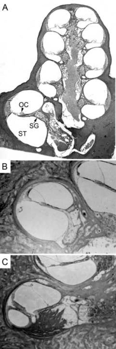
Figure 2.
2A. Anatomical section of guinea pig cochlea showing the scala tympani (ST), the chamber in which cochlear implants are inserted, the Organ of Corti (OC) where the sensory hair cells are found and enervated by spiral ganglion processes, and the spiral ganglion (SG) cell bodies. Electrical stimulation of the spiral ganglion processes or cell bodies can result in neuronal activation. 2B. Section of human cochlea with cochlear implant showing minimal tissue reaction. Small amounts of fibrous tissue were found around the electrode tip along the lateral wall. The implant had been in place for 5 years. 2C. Cochlear sections from another human patient with extensive new bone growth in the scala tympani between the electrode array and Rosenthal's canal, located at the right of the image. In addition, the osseous spiral lamina was fractured due to the implanted electrode. This cochlear implant had been in place for 8 years. Images 2B and 2C have been adapted from Kawano et al. (1998) with permission from Informa Healthcare.

