
Figure 1.
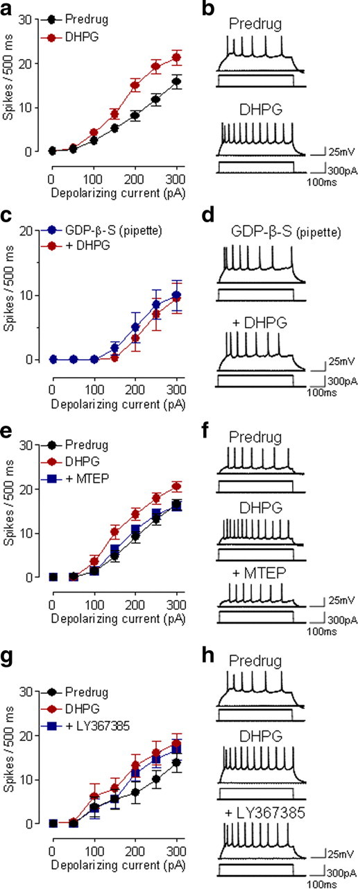
Group I mGluR5 activation increases excitability of CeLC neurons. a, DHPG (10 μm, 10–15 min) increased the input–output function of neuronal excitability of CeLC neurons (n = 31) significantly (p < 0.0001, main effect of drug, two-way ANOVA). Symbols show mean (± SE) number of spikes per 500 ms calculated from recordings like those in b. Action potentials were generated by depolarizing current injections of increasing magnitude (50 pA steps). b, Individual traces of action potentials generated by intracellular current injections (300 pA, 500 ms) before and during DHPG (10 μm). c, DHPG had no significant effect on neuronal excitability when a G protein inhibitor (GDP-β-S, 1 mm) was included in the patch pipette (n = 4 neurons; p > 0.05, two-way ANOVA). d, Individual example of action potentials evoked in a CeLC neuron with GDP-β-S included in the patch pipette (10 min after rupturing the cell membrane) and during the addition of DHPG. e, MTEP (1 μm, 15 min) decreased the facilitatory effect of DHPG (10 μm) significantly (n = 6 neurons; p < 0.0001, main effect of drug, two-way ANOVA). f, Action potentials evoked in a CeLC neuron in control (predrug), during DHPG, and during combined application of DHPG and MTEP. g, LY367385 (10 μm) had no significant effect on increased excitability induced by DHPG (10 μm) (n = 5 neurons; p > 0.05, main effect of drug, two-way ANOVA). h, Action potentials evoked in a CeLC neuron in control (predrug), during DHPG, and during combined application of DHPG and LY367385. a–g, DHPG, MTEP and LY367385 were superfused onto the brain slices. See Results for details of the statistical analysis.