
Figure 6.
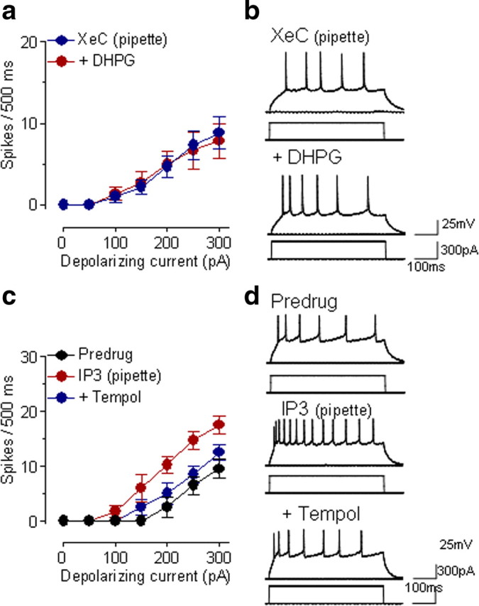
IP3 links group I mGluRs to ROS. a, In the presence of an intracellularly applied IP3 receptor blocker (XeC, 1 μm, included in the patch pipette) superfusion of DHPG (10 μm) onto the brain slices had no significant effect on neuronal excitability (n = 6 neurons; p > 0.05, main effect of drug, two-way ANOVA). Symbols show mean ± SE (see Fig. 1). b, Individual example of action potentials generated in a CeLC neuron by intracellular current injections (300 pA, 500 ms) before and during DHPG (10 μm, applied by superfusion) while XeC was injected into the cell. c, Intracellular application of IP3 (1 mm in the patch pipette) increased action potential firing significantly (n = 5 neurons; p < 0.0001, main effect of drug, two-way ANOVA). Superfusion of tempol (5 mm) onto the brain slices inhibited the facilitatory effect of IP3 significantly (n = 5 neurons; p < 0.0001, main effect of drug, two-way ANOVA). Action potential firing was measured immediately after whole-cell configuration was obtained, 10 min after rupturing the membrane, and during application of tempol for 10 min. d, Individual example of a CeLC neuron. See Results for details of the statistical analysis.