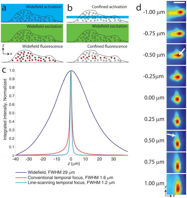
Figure 1.
Experimental considerations for 3D PALM. (a) Widefield activation (top panel) and excitation (middle panel) for localizing molecules (circles) throughout the entire cellular volume (bottom panel, filled circles). (b) Confined activation with widefield excitation for localization. (c) Sectioning performance of different illumination modalities. Integrated axial response of ~800 nm thick quantum dot film, moved through 80 μm axial range in 100 nm steps.Fluorescence was integrated over a 25 μm by 25 μm area, yielding the axial response under widefield (488 nm, blue), conventional- (800 nm, red), and line-scanning- temporal focus (800 nm, cyan) illumination. The latter shows reduced full width at half maximum (FWHM) compared to other excitation configurations, and reduced ‘tails’ at z positions further from focus. (d) Images of a 100 nm gold bead at different axial positions. The PSF shape is only coarsely approximated by an elliptical Gaussian function, as aberrations cause the intensity center-of-mass of the PSF to vary axially and ‘tails’ to vary asymmetrically (arrows). Scale bar 1 μm.

